If the published model available from the wwPDB is what you want, there is no reason to get into the complications of using a ".pdb" file; simply specify the wwPDB 4-character accession code.The user is responsible for the contents of files that FirstGlance does not fetch directly from the wwPDB using a 4-character accession code. Such files should strictly obey the PDB format: chain names must be single characters A-Z, a-z, 0-9; maximum 62 chains; maximum 99,999 atoms; etc.
COMPND MOL_ID: 1; COMPND 2 MOLECULE: PROTEIN; COMPND 3 CHAIN: A, B; EXPDTA ELECTRON MICROSCOPY REVDAT 1 REMARK 3 REFINEMENT.
(Technical: FirstGlance version 4.1 "peeks" into files with names ending ".pdb" [see dotPDB] that are on the same server as FirstGlance [all uploaded files] to find out whether they are ConSurf files. This is done before loading the PDB file. See peekConsurf() in moldoc.js, and ajaxThenJSmol() in scripts.js.)Unpublished models: ConSurf analysis: An unpublished model can be uploaded to the ConSurf server for analysis. The PDB file produced by ConSurf can then be uploaded to FirstGlance. FirstGlance will color the model by evolutionary conservation regardless of whether the filename contains "consurf" (see Technical note above).
load =5np6.cif select on chain=A or chain=B or chain=C write "5np6-chains-ABC.pdb"There is no header section in the resulting PDB file: Display 5np6-chains-ABC.pdb in FirstGlance. Jmol writes CONECT records for most covalent bonds. These are voluminous and unnecessary, and confuse some versions of Jmol. They should be deleted with a plain text editor.
load =5np6.cif select on within(group, within(20.0, (:A,:B,:C))) # ":A" is a synonym for "chain=A". write 5np6-within20-ABC.pdb # 24,987 atoms.When displayed in FirstGlance (after deleting the CONECT records), residues that have been separated from others in their chain (by the 20 Å cutoff) behave as "ligands+" in FirstGlance. The 16S rRNA is chain D, and the 23S rRNA is chain Y. Use "Find.." in FirstGlance to find chain=Y (yellow halos). Then use "Hide.." to hide "Atoms with Halos". Center the junction between the tRNA and the nascent peptide. Notice that the ribosomal amino acid closest to this junction (Arg81 of chain k, 50S ribosomal protein L16) is ~14 Å away from Gly46 of nascent peptide chain C, too far away to be the peptidyl-transferase. (View as Sticks, checking "more detail" to make distance measurements. Click Distances/Angles in the Tools tab for how to measure.)
Clicking on an atom produces an identification
report in the Jmol rectangle, to the lower left of the molecule (see
snapshot at right), and also in the browser's status bar.
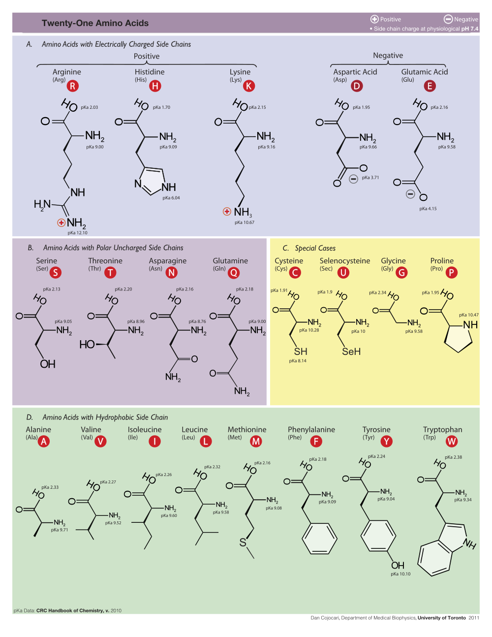
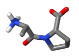
Amino acids are reported as
3-letter abbreviations,
and nucleotides as
|
CA = Carbon, Alpha (1st) CB = Carbon, Beta (2nd) CG, OG = Carbon/Oxygen, Gamma (3rd) CD = Carbon, Delta (4th) CE, NE, OE = Carbon/Nitrogen/Oxygen, Epsilon (5th) NZ = Nitrogen, Zeta (6th) NH = Nitrogen, Eta (7th) |
O3', O5' = 3' and 5' pentose oxygens C1' to C5' = 1' to 5' pentose carbons C2-C8, N1-N9, O2-O6 = elements in bases C5M = C5 Methyl (in Thymine) |
In addition to clicking, you can simply touch an atom with the mouse
(without moving the mouse, which is called "hovering"), and after a
delay of about two seconds, a yellow message will appear near the
mouse pointer (see snapshot at right) with an atom identification
report. This works best with Spin off.
These labels or marks are shown by default, but can be hidden by
unchecking the checkbox Labels: Show in the Focus Box at the bottom of any
of the upper left panels. (You may have to scroll the upper left panel
down to see this checkbox.)
|
| Biological Unit 1 of the Simian Virus 40 capsid (slabbed half) constructed from the crystallographic asymmetric unit and simplified to every 5th alpha carbon by FirstGlance in JSmol. |
| Reovirus Core 1ej6. 300 chains: 60 duplicates of the 5 chains in the asymmetric unit. | |||
|
|
|
|
| 1. Five Duplicated Chains | 2. Three Distinct Sequences | 3. Color by Distance | 4. Grayscale by Distance |
| Examples Illustrating Color Schemes for Large Biological Unit Assemblies | |||
| PDB ID | Chains in Biological Unit 1 | Chains in Asymmetric Unit | Sequence-Distinct Chains |
| 1ej6 | 300 | 5 | 3 |
| 3iyv | 216 | 18 | 2 |
| 1aym | 240 | 4 | 4 |
| 2GTL | 180 | 15 | 7 |
| 1a34 | 180 | 3 | 3 (Slab to see RNA inside!) |
| 6rd4 | 62 | 31 † | 18 |
| 2hhd | 4 | 4 | 2 |
|
|
Example: 3iyv, a 21 MDa clathrin cage 740 Å in diameter. The Molecule Information Tab reports that it has been simplified to every 8th alpha carbon. A more detailed view (every 2nd alpha carbon) is offered (see Option below), but provides few if any additional insights (see snapshots at right, colored by distance from center). See also an animation. In the Views tab, click Solid to see the Å values of the exaggerated radii of the alpha carbons, as well as other color schemes.Pros and Cons of Simplification:
In this group, Biological Unit 1 is identical with the deposited model, and no secondary
structure is specified:
|
Ala A Alanine
Arg R Arginine Asn N Asparagine Asp D Aspartic acid (mnemonic: asparDic) Cys C Cysteine |
Lys K Lysine (mnemonic: liKesine) Met M Methionine Phe F Phenylalanine (mnemonic: Fenylalanine) Pro P Proline Pyl O Pyrrolysine* |
mnemonic:
A Ala B Asx† C Cys D Asp asparDic E Glu gluEtamine F Phe Fenylalanine G Gly H His I Ile J K Lys liKesine L Leu M Met |
mnemonic:
N Asn asparagiNe O Pyl* P Pro Q Gln Quetamine R Arg aRginine S Ser T Thr U Sec* V Val W Trp tWptophan X Unk† Y Tyr tYrosine Z Glx† |
|
Gln Q Glutamine (mnemonic: Quetamine) Glu E Glutamic acid (mnemonic: gluEtamic) Gly G Glycine His H Histidine Ile I Isoleucine Leu L Leucine |
Sec U Selenocysteine* (mnemonic: seleniUm) Ser S Serine Thr T Threonine Trp W Tryptophan (mnemonic: tWyptophan) Tyr Y Tyrosine Val V Valine |
||
† Asx: Asn or Asp; Glx: Gln or Glu; Unk: unknown. IUPAC/IUB 1971 publication defining one-letter codes. | |||
| Resolution, Å | Interpretation |
|---|---|
| ≲1.7 | Most atoms very clearly seen. |
| ~1.7 - 2.2 | Many atoms clearly seen. |
| ~2.2 - 2.8 | Shapes of larger sidechains seen. |
| ~2.8 - 3.5 | Secondary structure & bulky groups seen. Atoms unclear. |
| ~3.5 - 4.0 | Most chains traceable. Atoms unclear. |
| ~4.0 - 6.0 | Some chains traceable. Atoms unclear. |
| ~6.0 - 8.0 | Shapes of domains at best. Atoms unclear. |
| ≳ 8.0 | Largest features seen roughly. Atoms unclear. |
| Resolution | Free R | ||
| GoodQ | Median | BadQ | |
| Free R Value | Grade | |
| <= (GoodQ - 0.02) | MUCH BETTER THAN AVERAGE at this resolution | |
| ------ GoodQ (best 25%) | > (GoodQ - 0.02) and <= ((GoodQ + Median)/2) | BETTER THAN AVERAGE at this resolution |
| ------ Median | > ((GoodQ + Median)/2) and <= ((Median + BadQ)/2) | AVERAGE at this resolution |
| ------ BadQ (worst 25%) | > ((Median + BadQ)/2) and <= (BadQ + 0.02) | WORSE THAN AVERAGE at this resolution |
| > (BadQ + 0.02) | UNRELIABLE | |
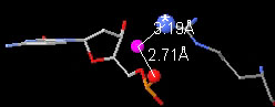
Water bridge in 1D66 between the sidechain
nitrogen of Arg 46
(chain B)
and a phosphate
oxygen of Arg 46
in Cytosine 13 (chain D).
|
appears below the molecule when any of the Unusual components or features
listed below are present.
halos
ALL UNUSUAL components or features
in the list below.
(Or enter ~all_unusual in the Find slot.)
below the molecule.
Rationale: According to the rules for PDB files, all atoms that are not members of standard residues in protein or nucleic acid chains should be designated "hetero". Hetero atoms are typically further subdivided: there is "solvent" (water and some inorganic anions such as sulfate and phosphate), and everything else is "ligand". Rare PDB files don't follow the rules, and these confuse Jmol. The result is atoms that are neither protein, nucleic acid, nor hetero. Within FirstGlance, these are deemed anomalous atoms.
Problem. FirstGlance will often fail to display
anomalous atoms appropriately. Some anomalous atoms could be invisible
in all views except Vines (with details on).
<form method='post' enctype='multipart/form-data'
action='https://proteopedia.org/cgi-bin/fgij/'>
Upload a PDB file of your own:
<br><small>
Uploaded data are https-encrypted..
</small><br>
<input type='file' name='file' size='50'>
<input type='submit' value='View in FirstGlance'>
</form>